Packing for College – Freshman Year

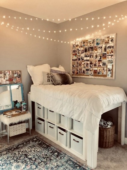
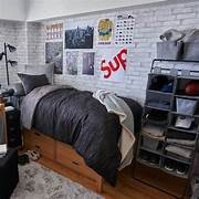
If you are moving into a dorm room, be prepared for some changes from home. Your dorm room will be a double, triple, or quad. If you are very lucky you will get a single room. Single rooms are usually reserved for those with special needs. In your dorm room, you will have a place for desks, closets, and beds. You might have a sink to use. Usually, everyone on the floor shares a communal bathroom/shower. The rooms are small and you will need to prioritize what you bring with you. This article helps you start to think about what to pack, Prepscholar.com has published a printable packing list as well here.

For meals, most four-year colleges have meal plans in communal dining halls. There are usually all types of food choices available. However, your dining hall will have specific open hours, and cannot adjust for every dietary need.

Be sure to bring your driver’s license, immunization card, health insurance card, list of medications, and past medical history. It is time for you to know these things. Snap a photo of the front (and back) of your cards and keep them saved in your favorites folder. You never know when you will need these things. Most children don’t know if they had chickenpox or if they had the chickenpox vaccination, so now is the time to get familiar with your medical history. Don’t forget your bank card and social security card.

It is a good idea to check and see what items the dorm will NOT allow. Things like:

Candles
Hookahs and electric cigarettes
Cooking appliances (except a microwave)
Air conditioners
Banners, tapestries, and flags
Hanging decorative lights
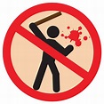
Weapons
Propane, gasoline, and lighter fluid
Lamps with plastic or cloth lampshades
Fog machines
Electric blankets

Other dorm room essentials (check with roommates about items that will be shared like microwave and minifridge – there will not be room for 3 microwaves and 3 minifridge’s in the dorm room).

Twin XL sheets (one set with pillowcases)Towels & washcloths
BlanketShower caddy to take to shower room
ComforterShower shoes
Mattress pad/coverShampoo, conditioner, soap/body wash
Under the bed storage boxesHair products
Laundry basket/bagComb or brush
Clothes hangersShaving supplies
Trash canCosmetics
Trash bagsToothpaste and toothbrush
Reading lightQ-tips/cotton balls
Minifridge (if not provided)Toilet paper/tissues or Kleenex
Microwave (if not provided)Nail clippers
2 cups, 2 plates, 2 bowls, 2 sets of silverwareLaundry detergent
Coffee mugDryer sheets
Reusable water bottleDish soap
Fan if you get hotHand soap
Rolling storage unitStorage for closet
Handheld chargeable vacuumBathrobe
Umbrella and raincoat/rain bootsCleaning supplies
Iron and ironing board (the little mini size) 

First Aid and Health Supplies

Painkillers/fever reducer Tylenol/ MotrinMedical tape
Band-aids all sizesGauze
NeosporinDisinfectant
CortisoneTweezers
Ace bandageCough and cold medicine
Birth control pills/condoms/other birth control 

School Supplies

NotebooksDesk organizerLaptop and laptop case
Planner*Desk lampNoise-canceling headphones
FoldersStamps/envelopesHDMI cable
Index cardsRubber bandsBackup chargers
Pens and pencilsPaper clipsAA and AAA batteries
HighlightersBinder clips1-2 power strips
SharpiesStaplercalculator
Post-itsBackpack or book bag 

Starter Grocery List

Ramen noodles & bowl to cook in the microwave
Microwavable soup packets or bowls
Snack packs (pretzels, almonds, trail mix, beef jerky)
Granola bars
Popcorn
Coffee or hot chocolate (whichever you like)
PB & J

Clothing

First, wait to bring your winter clothing, unless you are moving way up north. Choose a minimal wardrobe, plan for a tiny closet, and pack the most comfortable clothing. You will be walking around campus a lot, so pack at least one pair of sturdy walking shoes, rain boots, and an umbrella. Pack at least one but no more than two dressy outfits including shoes (something suitable for a job interview).

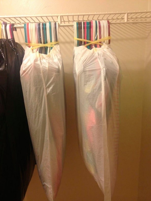
If you keep your clothes on their hangers then it is easier to transfer from the car to the closet. One space-saving tip is to use vacuum bags to pack towels and bedding (already washed). You can reuse these bags to store items in your closet that you do not use every day.  

Coordinate your clothing is more neutral colors that will match most items in your closet, this will go a long way to keeping your clothing to a minimum. You will also likely purchase a few items of clothing in the school colors while you are in school, so leave room for those. Don’t forget:

UnderwearLong sleeve shirts, sweatshirts
Socks, stockingsWorkout clothing
Jeans, pants, sweatpants1-2 outfits of professional attire
PJs (only need 2 or 3 pair)Belt
Shorts, skirtsSwimsuit
t-shirtsLight jacket, raincoat
Walking shoes, casual shoes, rain bootsSunglasses
Flip/flops or shower shoesWatch, hat, scarf

Problem-Based Learning

Problem-Based Learning (PBL) is an instructional approach that focuses on engaging students in solving real-world problems or complex scenarios. It is a student-centered, active learning methodology that promotes critical thinking, problem-solving skills, collaboration, and self-directed learning. Here are the key features and benefits of Problem-Based Learning:

Key Features of Problem-Based Learning:

  1. Real-world problems: PBL presents students with authentic, open-ended, and complex problems that reflect real-world situations. These problems are often multidisciplinary and require students to integrate knowledge from various subjects to find solutions.
  2. Student-centered approach: PBL places students at the center of the learning process. Instead of passive recipients of information, students become active participants who drive their learning by identifying knowledge gaps, conducting research, and seeking solutions.
  3. Collaboration and teamwork: PBL encourages collaborative learning and teamwork. Students work in small groups, sharing ideas, perspectives, and expertise to analyze problems, develop strategies, and reach consensus on potential solutions. Collaboration enhances communication, interpersonal skills, and the ability to work effectively in a team.
  4. Problem-solving skills: PBL emphasizes the development of critical thinking and problem-solving skills. Students learn to analyze complex problems, break them down into manageable components, identify relevant information, and propose solutions based on logical reasoning and evidence.
  5. Self-directed learning: PBL fosters self-directed learning skills. Students take responsibility for their learning process, set learning goals, conduct independent research, and explore resources to acquire the necessary knowledge and skills to address the problem at hand.
  6. Facilitator role of the teacher: In PBL, the teacher takes on the role of a facilitator or a guide rather than a lecturer. The teacher provides support, guidance, and resources to help students navigate the problem-solving process, clarify concepts, and develop critical thinking abilities.

Benefits of Problem-Based Learning:

  1. Active engagement: PBL promotes active engagement and deep learning. Students are actively involved in problem analysis, research, information synthesis, and the application of knowledge to propose solutions. This hands-on approach increases student motivation and interest in the subject matter.
  2. Application of knowledge: PBL allows students to apply theoretical concepts to real-world situations, making the learning experience more meaningful and relevant. It helps students bridge the gap between theory and practice, enhancing their understanding and retention of knowledge.
  3. Critical thinking and problem-solving skills: PBL cultivates critical thinking skills, such as analyzing, evaluating, and synthesizing information, as well as problem-solving skills, including generating creative solutions and making informed decisions. These skills are transferable and valuable beyond the classroom.
  4. Collaboration and communication skills: PBL encourages collaboration, effective communication, and teamwork. Students learn to work cooperatively, respect diverse perspectives, and effectively communicate their ideas, fostering interpersonal skills essential for future professional and personal contexts.
  5. Self-directed and lifelong learning: PBL nurtures self-directed learning skills, including research, information literacy, and the ability to set and achieve learning goals. These skills prepare students for lifelong learning, enabling them to adapt to new challenges and acquire knowledge independently.
  6. Higher-order thinking: PBL promotes higher-order thinking skills, such as analysis, synthesis, evaluation, and application. Students engage in complex problem-solving, requiring them to think critically, make connections across disciplines, and demonstrate a deeper understanding of the subject matter.
  7. Long-term retention: Due to the active and contextual nature of PBL, students often retain knowledge and skills acquired through problem-solving experiences for longer periods. The meaningful application of knowledge in solving real-world problems enhances memory consolidation and retrieval.

Examples of Problem-Based Learning

  1. Case study: A nursing student is presented with a case study involving a patient with multiple chronic conditions, such as diabetes, hypertension, and obesity. The student is tasked with developing a comprehensive care plan that addresses the patient’s physical health, medication management, lifestyle modifications, and psychological well-being.
  2. Ethical dilemma: A group of healthcare students, including nursing students, is presented with an ethical dilemma, such as the allocation of scarce resources during a pandemic. They must analyze the situation, consider ethical principles, and propose a fair and just approach to resource allocation.
  3. Community health project: Nursing students are assigned to a specific community and asked to identify a prevalent health issue affecting the population. They must conduct a needs assessment, develop an intervention plan, and propose strategies to improve the health outcomes of the community, considering factors such as cultural sensitivity, access to care, and health promotion.
  4. Interprofessional simulation: Nursing students, along with students from other healthcare disciplines, participate in a simulated patient scenario. The scenario involves a complex medical case where each student represents their profession and must collaborate to assess the patient, make clinical decisions, and develop a coordinated care plan.
  5. Quality improvement project: Nursing students are tasked with identifying a quality improvement opportunity within a healthcare setting, such as reducing hospital-acquired infections or medication errors. They must collect and analyze data, identify root causes, and propose evidence-based strategies to improve patient safety and outcomes.
  6. Global health challenge: Nursing students are presented with a global health challenge, such as addressing the high prevalence of a specific disease in a low-resource country. They must research the cultural, social, and economic factors influencing the disease, propose strategies for prevention and treatment, and consider the sustainability of the interventions.

Problem-Based Learning offers a learner-centered approach that develops critical thinking, problem-solving abilities, collaboration, and self-directed learning skills. By immersing students in authentic problem-solving experiences, PBL facilitates a deeper understanding of the subject matter and prepares students for the challenges they may face in their future careers. It promotes active engagement, application of knowledge, and the development of essential skills that are highly valued in the workforce, such as critical thinking, collaboration, and effective communication.

Furthermore, PBL fosters a positive and supportive learning environment where students feel empowered to take ownership of their learning and explore their interests. It encourages curiosity, inquiry, and the exploration of multiple perspectives, leading to a richer and more comprehensive understanding of the problem at hand.

PBL also helps students develop important meta-cognitive skills, such as self-reflection and self-assessment. They learn to evaluate their own progress, identify areas for improvement, and adjust their learning strategies accordingly. This metacognitive awareness enhances their ability to monitor and regulate their own learning, promoting lifelong learning habits.

Moreover, PBL can contribute to the development of transferable skills beyond subject-specific knowledge. Students learn to adapt to new situations, think critically and creatively, work effectively in teams, and communicate their ideas with clarity and confidence. These skills are vital in today’s rapidly changing world, where the ability to adapt, innovate, and collaborate is highly valued.

Overall, Problem-Based Learning offers numerous benefits, including active engagement, application of knowledge, development of critical skills, and preparation for real-world challenges. By immersing students in authentic problem-solving experiences, PBL not only deepens their understanding of the subject matter but also equips them with the skills and mindset necessary for success in their academic, professional, and personal lives.

References

Ghani, A. S. A., Rahim, A. F. A., Yusoff, M. S. B., & Hadie, S. N. H. (2021). Effective learning behavior in problem-based learning: a scoping review. Medical Science Educator31(3), 1199-1211.

Malmia, W., Makatita, S. H., Lisaholit, S., Azwan, A., Magfirah, I., Tinggapi, H., & Umanailo, M. C. B. (2019). Problem-based learning as an effort to improve student learning outcomes. Int. J. Sci. Technol. Res8(9), 1140-1143.

Moallem, M., Hung, W., & Dabbagh, N. (Eds.). (2019). The Wiley handbook of problem-based learning. John Wiley & Sons.

Trullàs, J. C., Blay, C., Sarri, E., & Pujol, R. (2022). Effectiveness of problem-based learning methodology in undergraduate medical education: a scoping review. BMC medical education22(1), 104.

The Pathophysiology and Risks of Heart Disease

Understanding and Mitigating Cardiac Risks

Heart disease, a term encompassing various cardiovascular conditions, remains a leading cause of mortality worldwide. Understanding the pathophysiology, risks, and preventive measures is crucial for managing and reducing cardiac risks. This article delves into the mechanisms of heart disease, the risks associated with it, and effective strategies to mitigate these risks. Additionally, it addresses the critical indicators of when to seek medical attention.

Pathophysiology of Heart Disease

Heart disease primarily involves the impairment of the heart’s ability to function effectively, often due to atherosclerosis, hypertension, or other cardiovascular abnormalities.

Atherosclerosis

Atherosclerosis is the buildup of fatty deposits, or plaques, within the arterial walls. These plaques narrow the arteries, reducing blood flow and oxygen supply to the heart muscle. Over time, this can lead to coronary artery disease (CAD), which may result in angina (chest pain) or myocardial infarction (heart attack).

Hypertension

Chronic high blood pressure, or hypertension, forces the heart to work harder to pump blood, leading to left ventricular hypertrophy (thickening of the heart muscle). Over time, this increased workload can weaken the heart, resulting in heart failure.

Other Cardiovascular Conditions

Other conditions such as valvular heart disease, arrhythmias, and cardiomyopathies also contribute to heart disease. These conditions disrupt the normal functioning of the heart, affecting blood flow and leading to various cardiac complications.

Risks of Heart Disease

Several risk factors contribute to the development and progression of heart disease. These can be categorized into modifiable and non-modifiable factors.

Non-Modifiable Risk Factors

  • Age: The risk of heart disease increases with age, particularly after 65.
  • Gender: Men are generally at higher risk of heart disease, though the risk for women rises and can surpass men’s after menopause.
  • Family History: A family history of heart disease significantly increases an individual’s risk.
  • Genetics: Certain genetic predispositions can elevate the risk of cardiovascular conditions.

Modifiable Risk Factors

  • Smoking: Tobacco use damages blood vessels and increases the risk of atherosclerosis and heart attacks.
  • High Blood Pressure: Uncontrolled hypertension strains the heart and arteries, leading to heart disease.
  • Cholesterol Levels: High levels of low-density lipoprotein (LDL) cholesterol contribute to plaque buildup in arteries.
  • Physical Inactivity: Sedentary lifestyles increase the risk of obesity, hypertension, and heart disease.
  • Poor Diet: Diets high in saturated fats, trans fats, and sodium can elevate heart disease risk.
  • Obesity: Excess body weight increases blood pressure, cholesterol levels, and the risk of diabetes, all of which are risk factors for heart disease.
  • Diabetes: Diabetes significantly increases the risk of cardiovascular complications due to high blood sugar levels damaging blood vessels.
  • Stress: Chronic stress can contribute to heart disease by increasing blood pressure and inflammation.

Reducing Cardiac Risk Factors

Implementing lifestyle changes and medical interventions can significantly reduce the risk of heart disease.

Healthy Diet

Adopting a heart-healthy diet is crucial. This includes:

  • Fruits and Vegetables: Consume a variety of fresh fruits and vegetables rich in vitamins, minerals, and antioxidants.
  • Whole Grains: Choose whole grains over refined grains for better heart health.
  • Lean Proteins: Opt for lean meats, fish, and plant-based proteins like beans and legumes.
  • Healthy Fats: Incorporate unsaturated fats from sources like olive oil, nuts, and avocados while reducing saturated and trans fats.
  • Limit Sodium: Reduce salt intake to manage blood pressure levels.

Regular Physical Activity

Engaging in regular exercise strengthens the heart and improves cardiovascular health. Aim for at least 150 minutes of moderate-intensity aerobic activity or 75 minutes of vigorous-intensity activity per week.

Smoking Cessation

Quitting smoking is one of the most effective ways to lower the risk of heart disease. Seek support from healthcare providers, support groups, or smoking cessation programs.

Weight Management

Maintaining a healthy weight through diet and exercise reduces the strain on the heart and lowers the risk of related conditions such as hypertension and diabetes.

Managing Stress

Adopt stress-reducing techniques such as mindfulness, meditation, yoga, or engaging in hobbies to lower stress levels and improve heart health.

Medical Management

For individuals with existing risk factors, medical interventions may be necessary. These include:

  • Medications: Drugs to manage blood pressure, cholesterol levels, and diabetes can help reduce cardiac risks.
  • Regular Check-ups: Routine medical examinations help monitor risk factors and ensure timely intervention.
  • Cardiac Rehabilitation: Structured programs that include exercise, education, and counseling to improve cardiovascular health.

When to Call the Doctor

Recognizing the signs of a heart problem and seeking prompt medical attention can be life-saving.

Symptoms to Watch For

  • Chest Pain or Discomfort: Especially if it spreads to the arms, back, neck, jaw, or stomach.
  • Shortness of Breath: Unexplained difficulty breathing during rest or activity.
  • Unexplained Fatigue: Persistent and unusual tiredness that interferes with daily activities.
  • Swelling: Edema in the legs, ankles, or feet, indicating heart failure.
  • Palpitations: Irregular or fast heartbeats that cause dizziness or lightheadedness.
  • Fainting: Sudden loss of consciousness.

Emergency Situations

Seek immediate medical attention if experiencing:

  • Severe chest pain or discomfort.
  • Sudden shortness of breath.
  • Weakness or numbness on one side of the body.
  • Difficulty speaking or understanding speech.
  • Loss of vision in one or both eyes.

Conclusion

Understanding the pathophysiology and risks of heart disease is essential for effective prevention and management. By adopting a healthy lifestyle, managing existing conditions, and knowing when to seek medical help, individuals can significantly reduce their risk of heart disease and improve their overall heart health.

Heart Sounds

Heart sounds, commonly heard during cardiac auscultation using a stethoscope, can provide valuable diagnostic information about the functioning of the heart. There are two main heart sounds, often labeled as S1 and S2, along with additional heart sounds that can indicate underlying cardiac conditions:

  1. S1 (Lub): This is the first heart sound, often described as a “lub” sound. It is caused by the closure of the mitral and tricuspid valves at the beginning of ventricular systole (when the ventricles contract to push blood out of the heart). S1 is associated with the onset of ventricular contraction and the beginning of the systolic phase.
  2. S2 (Dub): This is the second heart sound, often described as a “dub” sound. It is caused by the closure of the aortic and pulmonary valves at the end of ventricular systole (when the ventricles relax and begin to refill with blood). S2 marks the end of systole and the beginning of diastole.

In addition to S1 and S2, there are other heart sounds that may be heard under certain conditions:

  1. S3: This is a third heart sound, often indicative of heart failure. It occurs in early diastole during rapid passive filling of the ventricles. S3 is sometimes referred to as a “ventricular gallop” and is associated with conditions such as volume overload (e.g., in heart failure), mitral regurgitation, or ventricular septal defects.
  1. S4: This is a fourth heart sound, occurring late in diastole just before S1. It is often associated with reduced ventricular compliance, such as in conditions like hypertrophic cardiomyopathy or aortic stenosis. S4 is sometimes referred to as an “atrial gallop.”
  1. Murmurs: Murmurs are abnormal heart sounds caused by turbulent blood flow within the heart or blood vessels. They can indicate various cardiac conditions such as valve abnormalities (e.g., stenosis or regurgitation), septal defects, or abnormal flow patterns within the heart. Murmurs are often classified based on their timing (systolic, diastolic) and location (where they are heard best on the chest).
    • Aortic Stenosis (AS):
      • Timing: Typically systolic.
      • Intensity: Often crescendo-decrescendo (diamond-shaped) and may radiate to the carotid arteries.
      • Quality: Harsh or rough.
      • Location: Heard best at the right upper sternal border.
      • Associated clinical findings: Narrow pulse pressure, delayed and diminished carotid upstroke, possible ejection click.
  1. Mitral Regurgitation (MR):
    • Timing: Usually pansystolic.
    • Intensity: Varies, may be holosystolic or late systolic.
    • Quality: Soft blowing.
    • Location: Heard best at the apex and radiates to the axilla.
    • Associated clinical findings: S3 gallop, mitral valve prolapse, pulmonary hypertension, left atrial enlargement.
  1. Aortic Regurgitation (AR):
    • Timing: Early diastolic.
    • Intensity: High-pitched and blowing, may vary.
    • Quality: Decrescendo.
    • Location: Heard best at the left lower sternal border.
    • Associated clinical findings: Wide pulse pressure, water-hammer pulse (Corrigan’s pulse), diastolic murmur may have an associated Austin Flint murmur (mid-diastolic rumble at the apex).

  1. Mitral Stenosis (MS):
    • Timing: Mid-diastolic.
    • Intensity: May vary, often rumbling.
    • Quality: Low-pitched.
    • Location: Best heard at the apex with the bell of the stethoscope, often preceded by an opening snap.
    • Associated clinical findings: Loud S1, palpable presystolic thrill, left atrial enlargement, atrial fibrillation, possible pulmonary hypertension.
  1. Tricuspid Regurgitation (TR):
    • Timing: Often pansystolic.
    • Intensity: Varies, may be soft or blowing.
    • Quality: Holosystolic.
    • Location: Best heard at the left lower sternal border.
    • Associated clinical findings: Often secondary to right ventricular dilation or pulmonary hypertension, hepatic pulsations, jugular venous distention.
  1. Ventricular Septal Defect (VSD):
    • Timing: Usually pansystolic.
    • Intensity: Loud and harsh.
    • Quality: Holosystolic.
    • Location: Variable but often best heard at the left lower sternal border.
    • Associated clinical findings: Often heard in children with congenital heart disease, may have a palpable thrill, signs of heart failure.

Now put everything together

Interpreting heart sounds requires a combination of understanding the normal physiology of the heart and recognizing abnormal patterns associated with specific cardiac conditions. It’s essential for healthcare professionals to have thorough training and experience in cardiac auscultation to accurately diagnose and manage heart conditions.

Dating Violence: What Do I Need To Know?

Dating in adolescence and young adulthood is a crucial part of youth development, but it is also a period when many first experience relationship abuse. Teen dating violence (TDV) is a significant issue in the United States, affecting millions of teens each year.

TDV includes a range of controlling or aggressive behaviors such as psychological, physical, and sexual violence, stalking, bullying, and even homicide. Studies indicate that TDV affects between one in four to one in eight middle-to-high school students before age 18, with prevalence peaking between ages 16 and 18. The Youth Risk Behavior Surveillance Survey (YRBSS) reports that one in eight high school students experience physical or sexual dating violence, and many also face harassment and online bullying (Basile et al., 2020).

Sociodemographic factors play a role in the risk of dating violence. Older youth, ethnic/racial minority females, and those from low-income communities report higher rates of physical and sexual dating violence. Female students, LGBTQ+ students, and those unsure of their sexual identity have the highest prevalence of dating violence (Breidieg et al., 2014). The consequences of TDV are severe and long-lasting, including risky sexual behaviors, substance use, unintended pregnancies, eating disorders, mental health issues, and poor academic and social outcomes. TDV is also linked to future violence and chronic health problems (Coulter et al., 2017).

Rural youth report higher rates of physical dating violence compared to urban youth, yet they are understudied. Rural young males and females report higher rates of dating violence than their urban and suburban counterparts. This disparity highlights the unique challenges faced by rural youth, including limited access to support and accountability systems. Rural male perpetrators are significantly more likely than their urban counterparts to inflict serious bodily harm and use lethal weapons during abusive episodes. They are also twice as likely to threaten to murder an intimate partner, highlighting the severity of rural intimate partner violence (IPV; Huntley et al, 2019).

Rural victims face the worst psychosocial, mental, and physical outcomes due to violence-tolerant attitudes, negative bystander behaviors, and the assumed privacy of family violence in isolated communities. Additional regional risk factors include traditional gender norms, lack of confidential resources, long distances to care, and geographical isolation, making rural partner violence particularly distressing. Economic hardship, service provision gaps, the opioid epidemic, a shortage of trained service providers, and high social cohesion exacerbate rural dating violence. Cultural factors promoting self-sufficiency and trauma fatalism further prevent help-seeking, especially among rural men who have sex with men (MSM). These issues contribute to an aversion to help-seeking and abuse disclosure among rural boys and men (Hiebert, 2018).

Efforts to engage men and boys in violence reduction through gender transformative strategies have shown promising results, fostering gender equality and reducing violence. However, research on the impact of rurality and male-centered programs on dating violence among rural youths is limited. The COVID-19 pandemic has disrupted service delivery for IPV victims and perpetrators, particularly in underserved rural areas. Technology-based interventions offer potential solutions, providing privacy, convenience, and confidentiality. These interventions, such as smartphone apps and web-based platforms, have expanded support services for survivors but have not been widely studied with rural and male cohorts (Glass et al., 2022).

References

Basile KC, Clayton HB, DeGue S, et al. Interpersonal Violence Victimization Among High School Students — Youth Risk Behavior Survey, United States, 2019. MMWR Suppl 2020;69(Suppl-1):28–37. DOI: http://dx.doi.org/10.15585/mmwr.su6901a4

Breiding, M. J., Smith, S. G., Basile, K. C., Walters, M. L., Chen, J., & Merrick, M. T. (2014). Prevalence and characteristics of sexual violence, stalking, and intimate partner violence victimization–national intimate partner and sexual violence survey, United States, 2011. Morbidity and mortality weekly report. Surveillance summaries (Washington, D.C. : 2002)63(8), 1–18.

Coulter, R. W. S., Mair, C., Miller, E., Blosnich, J. R., Matthews, D. D., & McCauley, H. L. (2017). Prevalence of Past-Year Sexual Assault Victimization Among Undergraduate Students: Exploring Differences by and Intersections of Gender Identity, Sexual Identity, and Race/Ethnicity. Prevention science : the official journal of the Society for Prevention Research18(6), 726–736. https://doi.org/10.1007/s11121-017-0762-8

Glass, N. E., Clough, A., Messing, J. T., Bloom, T., Brown, M. L., Eden, K. B., Campbell, J. C., Gielen, A., Laughon, K., Grace, K. T., Turner, R. M., Alvarez, C., Case, J., Barnes-Hoyt, J., Alhusen, J., Hanson, G. C., & Perrin, N. A. (2022). Longitudinal Impact of the myPlan App on Health and Safety Among College Women Experiencing Partner Violence. Journal of interpersonal violence37(13-14), NP11436–NP11459. https://doi.org/10.1177/0886260521991880

Huntley, A. L., Potter, L., Williamson, E., Malpass, A., Szilassy, E., & Feder, G. (2019). Help-seeking by male victims of domestic violence and abuse (DVA): a systematic review and qualitative evidence synthesis. BMJ open9(6), e021960. https://doi.org/10.1136/bmjopen-2018-021960

Hiebert, B., Leipert, B., Regan, S., & Burkell, J. (2018). Rural Men’s Health, Health Information Seeking, and Gender Identities: A Conceptual Theoretical Review of the Literature. American journal of men’s health12(4), 863–876. https://doi.org/10.1177/1557988316649177

Respiratory Symptoms

Respiratory symptoms in respiratory illnesses encompass a broad spectrum of manifestations that significantly impact patients’ quality of life and functional status. Chronic respiratory conditions such as chronic obstructive pulmonary disease (COPD), asthma, interstitial lung disease (ILD), and cystic fibrosis (CF) often present with common symptoms such as dyspnea (shortness of breath), cough, wheezing, and chest tightness.

Dyspnea, commonly known as shortness of breath, is a complex symptom that arises from a wide range of physiological and pathological processes affecting the respiratory system, cardiovascular system, or both. The pathophysiology of dyspnea involves intricate interactions between sensory, neural, and muscular mechanisms.

  1. Sensory Receptors Activation: Dyspnea can be triggered by activation of sensory receptors located in the respiratory system, including mechanoreceptors, chemoreceptors, and nociceptors. These receptors detect changes in lung volume, oxygen and carbon dioxide levels, and tissue damage, respectively.
  2. Afferent Neural Pathways: Sensory signals from these receptors are transmitted via afferent neural pathways to the brainstem respiratory centers, including the medulla oblongata and pons, as well as higher cortical centers. These centers integrate and process sensory information, contributing to the perception of dyspnea.
  3. Activation of Respiratory Muscles: Dyspnea can also result from increased respiratory effort and work of breathing, which may be caused by conditions such as airway obstruction, lung hyperinflation, or respiratory muscle weakness. Increased respiratory effort leads to activation of accessory respiratory muscles, including the intercostal muscles and diaphragm, to overcome the underlying physiological derangements.
  4. Peripheral and Central Chemoreceptors: Chemoreceptors located in the peripheral and central nervous system play a crucial role in regulating respiratory drive by detecting changes in arterial blood gas levels, particularly oxygen and carbon dioxide concentrations. Hypoxemia and hypercapnia can stimulate these chemoreceptors, leading to increased respiratory rate and effort.
  5. Psychological and Emotional Factors: Dyspnea perception is also influenced by psychological and emotional factors, including anxiety, fear, and stress. These factors can amplify the sensation of dyspnea, even in the absence of significant physiological abnormalities.

Chronic cough is another prevalent symptom, frequently accompanied by sputum production, particularly in conditions like COPD and CF.

  1. Airway Irritation and Inflammation: Chronic cough frequently arises from irritation and inflammation of the airway mucosa. This inflammation can be triggered by a variety of factors, including viral or bacterial infections, allergens, environmental pollutants, and smoking. In response to these stimuli, inflammatory mediators such as histamine, prostaglandins, and leukotrienes are released, leading to airway hyperresponsiveness and increased cough reflex sensitivity.
  2. Mucociliary Dysfunction: Dysfunction of the mucociliary clearance mechanism in the respiratory tract can contribute to chronic cough. Normally, cilia lining the airway epithelium help to clear mucus and foreign particles from the airways. In conditions such as chronic bronchitis or cystic fibrosis, impaired ciliary function results in mucus accumulation, leading to cough as a mechanism to clear the airways.
  3. Sensory Neuropathy: Alterations in the cough reflex pathway and sensory neuropathy can contribute to chronic cough. Conditions such as postnasal drip syndrome, upper airway cough syndrome (formerly known as postnasal drip syndrome), and cough variant asthma involve heightened sensitivity of cough receptors in the upper airway or lower respiratory tract, leading to chronic cough.
  4. Psychogenic Causes: Psychological factors such as anxiety, stress, and psychogenic cough can also contribute to the development of chronic cough. These factors can modulate the central cough reflex pathway, resulting in persistent coughing even in the absence of underlying organic pathology.

Wheezing is a high-pitched, whistling sound produced during expiration and sometimes inspiration, which typically arises from narrowed or obstructed airways. The pathophysiology of wheezing involves a complex interplay of anatomical, physiological, and pathological factors within the respiratory system:

  1. Airway Obstruction: Wheezing often occurs due to partial obstruction of the airways, leading to turbulent airflow during breathing. Common causes of airway obstruction include bronchoconstriction, mucosal edema, inflammation, and the presence of excessive mucus.
  2. Bronchoconstriction: Constriction of the smooth muscle in the bronchioles, a hallmark feature of conditions like asthma and chronic obstructive pulmonary disease (COPD), narrows the airways and increases airway resistance. This bronchoconstriction results in airflow limitation and turbulent airflow, manifesting as wheezing.
  3. Mucosal Edema and Inflammation: Inflammatory conditions such as asthma, bronchitis, and respiratory infections can lead to swelling and inflammation of the airway walls. Mucosal edema narrows the airways, while increased mucus production further obstructs airflow, contributing to wheezing.
  4. Excessive Mucus Production: Conditions associated with excessive mucus production, such as chronic bronchitis, cystic fibrosis, and bronchiectasis, can cause mucus accumulation within the airways. This excess mucus obstructs airflow and contributes to wheezing sounds during breathing.
  5. Airway Hyperresponsiveness: Individuals with heightened airway reactivity, such as those with asthma, are more susceptible to airway narrowing and bronchoconstriction in response to various triggers. This increased airway responsiveness contributes to recurrent wheezing episodes, especially during exposure to allergens, irritants, or respiratory infections.
  6. Dynamic Airway Collapse: In conditions like tracheobronchomalacia, weakened or floppy airway walls can collapse during expiration, leading to partial airway obstruction and wheezing. Dynamic airway collapse is exacerbated during increased airflow velocity, such as during forced expiration, and can manifest as inspiratory or expiratory wheezing.

Tachypnea, defined as rapid breathing or an increased respiratory rate, can be a physiological response to various factors, including increased metabolic demand, hypoxia, acidosis, or fever. The pathophysiology of tachypnea involves alterations in respiratory drive, gas exchange, or lung mechanics, depending on the underlying cause:

  1. Respiratory Drive: Tachypnea can occur due to stimulation of the respiratory centers in the brainstem, which regulate breathing. Factors such as hypoxia, hypercapnia, metabolic acidosis, and pain can stimulate these respiratory centers, leading to an increase in respiratory rate as the body attempts to correct these abnormalities.
  2. Gas Exchange Abnormalities: Conditions that impair gas exchange in the lungs, such as pneumonia, pulmonary edema, or acute respiratory distress syndrome (ARDS), can result in hypoxemia. Hypoxemia triggers compensatory mechanisms to increase ventilation and improve oxygenation, leading to tachypnea.
  3. Lung Mechanics: Tachypnea can also result from alterations in lung mechanics, such as airway obstruction or decreased lung compliance. Airway obstruction, as seen in conditions like asthma, chronic obstructive pulmonary disease (COPD), or foreign body aspiration, increases airway resistance and requires increased respiratory effort, leading to tachypnea. Decreased lung compliance, as observed in conditions like pulmonary fibrosis or atelectasis, requires increased work of breathing to overcome stiff or collapsed lung tissue, resulting in tachypnea.
  4. Fever: Elevated body temperature, as seen in systemic infections or inflammatory conditions, can lead to tachypnea as part of the body’s response to increase heat loss through respiration. Fever stimulates the respiratory centers in the brainstem, leading to an increase in respiratory rate.
  5. Pain: Severe pain, particularly in the thoracic or abdominal region, can stimulate the respiratory centers and lead to tachypnea as a reflex response to alleviate discomfort or distress.

Crackles, also known as rales, are abnormal lung sounds characterized by discontinuous, brief, and non-musical sounds heard during inspiration or expiration. These sounds arise from the sudden opening of small airways, the movement of air through fluid-filled airways, or the popping open of collapsed alveoli. The pathophysiology of crackles can be attributed to various underlying mechanisms:

  1. Airway Secretions: Crackles can occur when air flows through narrowed or partially obstructed airways, causing the movement of secretions or mucus. In conditions such as bronchitis, pneumonia, or bronchiectasis, excessive mucus production or inflammation can lead to airway obstruction and the formation of crackles as air passes through the fluid-filled airways.
  2. Alveolar Instability: Crackles may also result from the sudden opening of collapsed alveoli or the recruitment of previously closed airways during inspiration. Conditions such as acute respiratory distress syndrome (ARDS), pulmonary fibrosis, or congestive heart failure (CHF) can lead to alveolar collapse or atelectasis, which can result in crackles upon re-expansion of the alveoli.
  3. Interstitial Fluid Accumulation: In conditions such as interstitial lung disease (ILD) or pulmonary edema, fluid accumulation within the interstitial spaces of the lungs can impair gas exchange and lead to crackles. As air moves through fluid-filled interstitial spaces during inspiration, crackles may be heard due to the disruption of normal airflow patterns.
  4. Airway Collapse: Dynamic airway collapse, as seen in conditions like tracheobronchomalacia or bronchial asthma, can result in intermittent narrowing of the airways during respiration. Crackles may occur as collapsed airways suddenly reopen during inspiration, leading to turbulent airflow and the production of crackling sounds.
  5. Peripheral Airway Closure: During expiration, crackles may arise from the sudden closure of peripheral airways. In conditions such as chronic obstructive pulmonary disease (COPD) or asthma, peripheral airway narrowing or bronchoconstriction can lead to premature airway closure during expiration, resulting in crackles upon subsequent inspiration.

Respiratory retractions, also known as intercostal retractions or subcostal retractions, are visible inward movements of the soft tissues between the ribs during inspiration. They typically indicate increased effort required to breathe due to airway obstruction, respiratory muscle fatigue, or decreased lung compliance. The pathophysiology of respiratory retractions involves the following mechanisms:

  1. Airway Obstruction: Respiratory retractions often occur in response to partial or complete airway obstruction, leading to increased resistance to airflow. Conditions such as asthma, bronchiolitis, or foreign body aspiration can cause narrowing or blockage of the airways, requiring increased respiratory effort to overcome the obstruction.
  2. Increased Airway Resistance: In conditions characterized by increased airway resistance, such as bronchospasm or bronchoconstriction in asthma or chronic obstructive pulmonary disease (COPD), the respiratory muscles must work harder to move air in and out of the lungs. This increased respiratory effort can result in visible retractions of the intercostal spaces or subcostal area during inspiration.
  3. Respiratory Muscle Fatigue: Prolonged or strenuous breathing against increased resistance can lead to fatigue of the respiratory muscles, including the diaphragm, intercostal muscles, and accessory muscles of respiration. As the muscles fatigue, they may become less effective in generating adequate airflow, leading to retractions as the body attempts to increase respiratory effort.
  4. Decreased Lung Compliance: Conditions that decrease lung compliance, such as pulmonary fibrosis or atelectasis, impair the ability of the lungs to expand and accommodate air during inspiration. This results in increased work of breathing and visible retractions as the respiratory muscles attempt to overcome the reduced lung compliance and expand the lungs.
  5. Increased Work of Breathing: Any condition that increases the work of breathing, such as hypoxemia, hypercapnia, or metabolic acidosis, can lead to visible retractions as the body attempts to compensate for the respiratory derangements. The increased respiratory effort required to maintain adequate gas exchange can result in retractions of the chest wall during inspiration.

Chest tightness, also referred to as chest discomfort or chest pressure, is a common symptom experienced in various respiratory conditions. The pathophysiology of chest tightness in respiratory conditions involves a combination of anatomical, physiological, and neurological factors:

  1. Airway Constriction: Chest tightness can result from bronchoconstriction, the narrowing of the airways due to the contraction of smooth muscle surrounding the bronchi and bronchioles. This occurs in conditions such as asthma, where triggers such as allergens or irritants lead to inflammation and bronchoconstriction, causing a sensation of tightness in the chest.
  2. Air Trapping and Hyperinflation: In chronic obstructive pulmonary disease (COPD), characterized by chronic bronchitis and emphysema, air trapping and hyperinflation of the lungs can contribute to chest tightness. Air trapping occurs when the small airways collapse during expiration, trapping air in the alveoli and leading to increased lung volume. This increased lung volume can exert pressure on the chest wall, resulting in a sensation of tightness.
  3. Inflammation and Edema: Inflammatory conditions affecting the respiratory tract, such as pneumonia or bronchitis, can lead to swelling and edema of the airway walls. This inflammation and edema can cause narrowing of the airways, increased airway resistance, and a sensation of chest tightness.
  4. Accumulation of Secretions: Excessive mucus production and the accumulation of secretions within the airways can contribute to chest tightness. Conditions such as bronchiectasis, cystic fibrosis, or chronic bronchitis are characterized by increased mucus production, leading to airway obstruction and chest tightness.
  5. Anxiety and Hyperventilation: Psychological factors such as anxiety or panic attacks can lead to hyperventilation, resulting in respiratory alkalosis and a sensation of chest tightness. Hyperventilation causes a shift in the acid-base balance of the blood, leading to decreased carbon dioxide levels and respiratory alkalosis, which can manifest as chest tightness or discomfort.

Stridor is a high-pitched, wheezing sound that occurs during breathing and is often associated with obstruction or narrowing of the upper airway. The pathophysiology of stridor involves a disruption in the normal airflow through the upper airway, leading to turbulent airflow and the production of sound. Several factors can contribute to the development of stridor:

  1. Airway Obstruction: Stridor typically occurs when there is partial obstruction or narrowing of the upper airway. This obstruction can be due to various causes, including inflammation, swelling, foreign bodies, tumors, or structural abnormalities such as congenital malformations or trauma.
  2. Inflammation and Swelling: Inflammatory conditions affecting the upper airway, such as croup, epiglottitis, or laryngotracheobronchitis, can cause swelling of the tissues lining the airway. This swelling narrows the airway and disrupts airflow, leading to the production of stridor.
  3. Congenital Anomalies: Structural abnormalities present at birth, such as laryngomalacia (floppy larynx), tracheomalacia (weakness of the tracheal cartilage), or vascular rings, can result in obstruction or narrowing of the upper airway. These anomalies can lead to turbulent airflow and the characteristic sound of stridor.
  4. Foreign Bodies: Inhalation of foreign objects, such as food particles, toys, or small objects, can obstruct the upper airway and cause stridor. The presence of a foreign body creates a physical barrier to airflow, leading to turbulent airflow and the production of sound.
  5. Tumors: Benign or malignant growths in the upper airway, such as laryngeal or tracheal tumors, can cause obstruction and lead to the development of stridor. Tumors can compress or invade the airway, disrupting airflow and causing turbulent flow.
  6. Neuromuscular Disorders: Conditions that affect the muscles or nerves controlling the upper airway, such as vocal cord paralysis or neuromuscular diseases like myasthenia gravis, can result in weakness or paralysis of the muscles involved in breathing. This weakness can lead to airway collapse or obstruction, contributing to the development of stridor.

Cyanosis occurs when there is an increased concentration of deoxygenated hemoglobin in the blood. Hemoglobin is the protein in red blood cells that carries oxygen from the lungs to the body’s tissues. When hemoglobin binds with oxygen, it forms oxyhemoglobin, which gives blood its bright red color. However, when hemoglobin is not fully saturated with oxygen, it appears bluish in color.

The pathophysiology of cyanosis involves one or more of the following mechanisms:

  1. Decreased Oxygenation of Blood: Cyanosis often occurs when there is insufficient oxygenation of the blood in the lungs. This can happen due to respiratory conditions such as pneumonia, asthma, chronic obstructive pulmonary disease (COPD), or hypoventilation (reduced breathing). In these conditions, inadequate gas exchange in the lungs results in a higher proportion of deoxygenated hemoglobin in the bloodstream.
  2. Circulatory Impairment: Cyanosis can also result from circulatory problems that affect the delivery of oxygen-rich blood to tissues. Conditions such as congenital heart defects, heart failure, shock, or peripheral vascular disease can impair blood flow or reduce oxygen delivery to tissues, leading to cyanosis.
  3. Shunting of Blood: In some cases, cyanosis can occur due to abnormal shunting of blood within the circulatory system. A shunt is a connection between two blood vessels or chambers of the heart that allows blood to bypass the lungs, where it would normally pick up oxygen. This can happen in congenital heart defects like tetralogy of Fallot or transposition of the great arteries, where blood is redirected from the lungs to the systemic circulation without being adequately oxygenated.
  4. Methemoglobinemia: Methemoglobin is a form of hemoglobin that cannot bind oxygen effectively. Normally, only a small percentage of hemoglobin is in the methemoglobin form. However, certain substances or medications can increase methemoglobin levels, leading to cyanosis. This condition is known as methemoglobinemia.

These respiratory symptoms not only impair patients’ daily activities but also contribute to anxiety, depression, and decreased quality of life. Effective management of chronic respiratory symptoms involves a multidisciplinary approach, including pharmacological interventions, pulmonary rehabilitation, patient education, and lifestyle modifications, tailored to individual patient needs and preferences. Early recognition and proactive management of respiratory symptoms are essential for optimizing patient outcomes and enhancing overall well-being in individuals living with chronic respiratory conditions.

Having that Difficult Conversation with Patients or Colleagues

pink flower on a collage background

Beautiful Dalia

There are always times when we need to have a difficult conversation. Perhaps it is with a friend, peer, colleague, or patient. The topic continuously varies. Many of us humans, who I’m referring to, may dread difficult conversations or conflict. We often learned to avoid difficult conversations as we grew up, probably because these conversations were not dealt with well by our parents or peers. Learning to deal with difficult conversations systematically can help everyone be better communicators and convey the message more positively. Remember, the principles of a positive discussion are the same.

Mary Jean is a coworker who has been coming to work late several times a week over the past month. You need to talk with her about coming to work on time.

Benjamin is a patient who has a terminal illness and wants to go to Hospice. His family disagrees and wants him to receive all treatment. You need to mediate a conversation between Benjamin and his family.

The Right State of Mind

Both examples are of possible problematic situations. So, how should you proceed? Let’s discuss some steps first to get in the right state of mind. It is essential to practice scenarios like this, not in person or out loud; practice in your head as you get ready for bed and relax for the evening. Practice all possible outcomes, from the worst possible to the best possible, and everything in between. Next, keep your state of mind positive. Whenever you practice scenarios, picture yourself calm, positive, and effective when communicating.

It would help if you held every conversation with empathy and compassion. Show the person you care about them and anything they are going through. You share compassion and empathy by listening carefully, being authentic, and genuinely caring.

  • Compassion is showing concern for someone who is suffering.
  • Compassion leads to an action to help others.
  • Compassion inspires positive feelings.
  • Empathetic persons feel the emotions of others.
  • Empathy leads to understanding.

Practice these over and over before the difficult conversation and even anytime you anticipate a difficult conversation. It does not matter who the conversation is supposed to be with, practice. Practice the right way to say something. Practice the unexpected as well as the expected. Don’t give yourself a script; follow a different script each time you practice. Use other words each time so that you are prepared for different scenarios and can adapt to anything that comes your way. This technique is called Guided Imagery and is extremely useful.

Breathing

Practice basic principles of mindful breathing. Mindful breathing is a form of meditation you can do when practicing different scenarios at bedtime or during other quiet periods. First, get in a relaxed pose. Any position that you are comfortable in will work. Take a slow, deep breath in, then release and exhale slowly until all air is out of your lungs. Do not take a break in between each breath. Practice this for 10 minutes for about a week, then move on to 20 minutes. Do remember that you can stop at any time. Feel yourself, be calm, be relaxed. Notice any areas where you are tense, and relax those muscles. As you breathe, tune into your breath and feel your breath move in and out. Feel yourself relax.

Now that you have practiced a bit, you will be more ready for the expected and the unexpected. You will be more prepared for that difficult conversation than ever. Now, let us move toward a systematic way to respond to someone and how to be relaxed and compassionate while you are responding.

The Systematic Approach

Start with an opening comment or question. Show empathy while you are at it. If you are delivering terrible news, a death, for example, you should try to ensure that they have someone to be with them or drive them to where they need to go. You might say

  • “I’m concerned about your attendance lately. Is everything okay at home?”
  • “I’ve asked you here today to talk about Benjamin’s wishes for his care.”
  • “It is nice to see you today; how have things been with you and your family lately.”
  • “Tell me what you know about Benjamin’s health.”

Take Time to Understand Their Perspective

You do not have to answer right away. Take several breaths, relax, and consider their perspective or issues. It is okay to say I’m so sorry that this happened to you. or that you are going through this. If they begin to cry, give them space and acknowledge the distress before moving on. Remember to deliver the news in a calm, compassionate manner. Do not try to convey the other person’s sympathy by saying things like I feel wrong about this, or This is hard for me to do.

Explain the Change in Their Behavior You Expect to See

Work-Related Issue

Explain the issue that you are having and what changes you expect to see. Do so compassionately and calmly. You cannot fix the situation for them, but you can provide suggestions if they would like suggestions. But first, ask them what they think the solution is. If they cannot come up with a solution, ask them if they would like you to provide suggestions. If they say no, respect their wishes.

Healthcare Issue

If you are discussing a medical issue with patients or family members, describe the issue at hand and how that issue impacts the patient and family. Explain medical words or procedures and give timelines when necessary. It is important to be nonjudgmental, reflective, and compassionate. Provide teaching materials if that helps. Use I statements or allow the patient to use I statements. Mediate situations where the patient needs to tell their family how they feel and what they want. Be there to support the patient in those instances. Then, give them a bit of a break to process. Just listen.

End the Session

To end these conversations, paraphrase the problem and the plan to move forward. If no plan was established, then ask them to reflect and get back to you. Give them a specific timeline of when you expect to hear from them. Be encouraging, calm, and empathetic. Then, follow up with them at the right time to complete the circle.

References

Bigelow, S., Medzon, R., Siegel, M., & Jin, R. (2024). Difficult Conversations: Outcomes of Emergency Department Nurse-Directed Goals-of-Care Discussions. Journal of Palliative Care, 39(1), 3–12. https://doi.org/10.1177/08258597221149402

Hannans, J., & Nevins, C. (2024). Using Mixed Reality to Practice Difficult Conversations. Clinical Simulation in Nursing, 86, N.PAG. https://doi-org.proxy.mul.missouri.edu/10.1016/j.ecns.2023.101472

King, C., & Williams, B. (2021). Enabling difficult conversations in the Australian health sector. Australian Journal of Advanced Nursing, 38(3), 5–13. https://doi-org.proxy.mul.missouri.edu/10.37464/2020.383.310

Sulistio, M. S., Chen, C. L., Eleazu, I., Godfrey, S., Abraham, R. A., & Toft, L. E. B. (2023). Personal Actions to Create a Culture of Inclusion: Navigating Difficult Conversations With Medical Colleagues. Annals of Internal Medicine, 176(11), 1520–1525. https://doi-org.proxy.mul.missouri.edu/10.7326/M23-1374